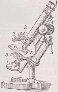
Microscopes are distinguished as simple or compound. In the former, the rays which enter the eye of the observer come from an object brought near to it after refraction through either a single lens or a combination of lenses acting as a single lens,—its action as a “magnifier” depending on its enabling the eye to form a distinct image of the object at a much shorter distance than would otherwise be possible. The latter consists of at least two lenses, so placed relatively to the object, to the eye, and to one another that an enlarged image of the object, formed by the lens placed nearest to it (the “object-glass ”), is looked at through the lens nearest the eye (the “eye-glass”), which acts as a simple microscope in “magnifying” it; so that the compound microscope may be described as a simple microscope used to look at an enlarged image of the object, instead of at the object itself....
History of the Simple Microscope.— Any solid or liquid transparent medium of lenticular form, having either one convex and one flat surface or two convex surfaces whose axes are coincident, may serve as a “magnifier,”—what is essential being that it shall have the power of so refracting the rays which pass through it as to cause widely diverging rays to become either parallel or but slightly divergent. Thus if a minute object be placed on a slip of glass, and a single drop of water be carefully placed upon it, the drop will act as a magnifier in virtue of the convexity of its upper surface; so that when the eye is brought sufficiently near it (the glass being of course held horizontally, so as not to distort the spherical curvature of the drop) the object will be seen much enlarged. And if a small hole be made in a thin plate of metal, and a minute drop of water be inserted in it, this drop, having two convex surfaces, will serve as a still more powerful magnifier. There is reason to believe that the magnifying power of transparent media with convex surfaces was very early known. A convex lens of rock-crystal was found by Layard among the ruins of the palace of Nimrud. And it is pretty certain that, after the invention of glass, hollow spheres blown of that material and filled with water were commonly used as magnifiers (comp. vol. xiv. p. 577). The perfection of gem-cutting shown in ancient gems, especially in those of very minute size, could not have been attained without the use of such aids to the visual power; and there can be little doubt that the artificers who could execute these wonderful works could also shape and polish the magnifiers best suited for their own or others’ use. Though it is impossible to say when convex lenses of glass were first made by grinding, it is quite certain that they were first generally used to assist ordinary vision as “spectacles,” the use of which can be traced back nearly six centuries; and not only were spectacle-makers the first to produce glass magnifiers (or simple microscopes), but by them also the telescope and the compound microscope were first invented. There seems no reason to believe, however, that lenses of very high magnifying power (or short focus) were produced until a demand for them had been created by the introduction of the compound microscope, in which such lenses are required as “object-glasses ”; and the difficulty of working lenses of high curvature with the requisite accuracy led in the first instance to the employment of globules made by fusing the ends of threads of spun glass. It was in this way that Robert Hooke shaped the minutest of the lenses with which he made many of the numerous discoveries recorded in his Micrographia∖ and the same method was employed by the Italian microscopist Father Di Torre. It seems to have been Leeuwenhoek that first succeeded in grinding and polishing lenses of such short focus and perfect figure as to render the simple microscope a better instrument for most purposes than any compound microscope then constructed,—its inferiority in magnifying power being more than counterbalanced by the superior clearness of the retinal picture. And, in despair of any such modification in the compound form as should remove the optical defects which seemed inherent in its plan of construction, scientific opticians and microscopic observers alike gave their chief attention for a considerable period to the improvement of the simple microscope. In order that the nature of these improvements may be understood, the principle of its action must be first explained.
The normal human eye has a considerable power of selfadjustment, by which its focal length is so varied that it forms equally distinct pictures of objects brought within ordinary reading distance (say 10 inches) and of objects whose distance is many times that length,—the size of the visual picture of any object diminishing, however, with the increase in the distance to which it is removed, and the amount of detail distinguishable in it following the same proportion. Thus a man who looks across the street at a placard posted on the opposite wall may very distinctly see its general form and the arrangement of its heading, and may be able to read what is set forth in its largest type, whilst unable to separate the lines, still more to read the words, of what is set forth below. But by crossing the street so as to bring his eye nearer the picture he finds himself able,to read the smaller type as easily as he before read the larger,—the visual picture on his retina having been magnified, say 10 times in linear dimension, by the reduction of the distance of his eye from 40 feet to 4. Similarly, if he holds a page of excessively minute type at arm’s length (say 40 inches) from his eye, he may be unable to read it, not because his eye does not form a distinct retinal picture of the page at that distance, but because the details of that picture are too minute for him to distinguish them. But if he brings the page from 40 inches to 10 inches distance, he may be able to read it without difficulty,—the retinal picture being enlarged four times linear (or sixteen times superficial) by this approximation. Now the rays that enter the eye from each point of a remote object diverge so little as to be virtually parallel; but the divergence increases with the approximation of the object to the eye, and at 10 inches the angle of their divergence is as wide as permits the ordinary eye to bring them to a focus on the ïetina. When the object is approximated more closely, an automatic contraction of the pupil takes place, so that the most diverging rays of each pencil are cut off, and a distinct picture may be formed (though not without a feeling of strain) when the object is (say) from 5 to 8 inches distant,—giving still greater minuteness of visual detail in conformity with the increase of size. A further magnifying power may be obtained without the interposition of any lens, by looking at an object, at 2 or 3 inches distance, through a pin-hole in a card; for by thus cutting off the more divergent rays of each pencil, so as to admit only those which can be made to converge to a focus on the retina at that distance, a distinct and detailed picture may be obtained, though at the expense of a great loss of light. Moreover, although an ordinary eye does not form a distinct picture of an object at less than from 10 to 6 inches distance, a “myopic” or “short-sighted” eye (whose greater refractive power enables it to bring rays of wider divergence to a focus on the retina) may form an equally distinct picture of an object at from 5 to 3 inches distance; and, as the linear dimensions of that picture will be double that of the preceding, the object will be “magnified” in that proportion, and its details more clearly seen.
The effect of the interposition of a convex lens between the eye and an object nearly approximated to it primarily consists in its reduction in the divergence of the rays of the pencils which issue from its several points, so that they enter the eye at the moderate divergence which they would have if the object were at the ordinary nearest limit of distinct vision. And, since the shorter the focus of the lens the more closely may the object be approximated to the eye, the retinal picture is enlarged, causing the object to appear magnified in the same proportion. Not only, however, are the component rays bf each pencil brought from divergence into convergence, but the course of the pencils themselves is changed, so that they enter the eye under an angle corresponding to that under which they would have arrived from a larger object situated at a greater distance; and thus, as the picture formed upon the retina by the small object ab, fig. 1, corresponds in all respects with that which would have been made by the same object AB of several times its linear dimension viewed at the nearest ordinary limit of distinct vision, the object is seen (by the formation of a “virtual image ”) on a magnified scale.
It is obvious that the “magnifying power” of any convex lens so used is measured by the ratio between the dimensions of the retinal picture formed with its assistance and those of the picture formed by the unaided eye. Thus, if by the use of a convex lens having 1 inch focal length we can form a distinct retinal image of an object at only an inch distance, this image will have ten times the linear dimensions of that formed by the same object at a distance of 10 inches, but will be only eight times as large as the picture formed when the object can be seen by ordinary vision at 8 inches distance, and only four times as large as the picture of the same object formed by a myopic eye at a distance of 4 inches. It is usual to estimate the magnifying power of single lenses (or of combinations that are used as such) by the number of times that their focal length is contained in 10 inches,—that of 1 inch focus being thus taken as ten times, that of τ ⅛ inch as one hundred times, and so on. But the rule is obviously arbitrary, as the actual magnifying power varies in each individual with the nearest limit of distinct vision. Thus for the myopic who . can see an object clearly at 4 inches distance, the magnifying powers of a 1 inch and τ 1 σ inch lens will be only 4 and 40 respectively. The amplifying power of every single convex lens, however, is impaired (1) by that inability to bring to the same focus the rays which fall upon the central and the marginal parts of its surface which is called “spherical aberration,” and (2) by that dispersion of the rays of different wave-lengths, in virtue of their different refrangibilities, which produces coloured fringes around the points and lines of the visual picture, and is therefore called “chromatic aberration” (see Light). These aberrations increase with the “angle of aperture” given to the lens, that is, with the proportion between the diameter of its actual “opening” and the focal distance of the object; and thus, when a single lens of very short focus is used in order to gain a high magnifying power, such a reduction of its aperture by a perforated diaphragm or “stop” becomes necessary (in order, by excluding the peripheral rays, to obtain tolerable “definition” with freedom from false colour) that the amount of light admitted to the eye is so small as only to allow the most transparent objects to be thus viewed, and these only very imperfectly. In order to remedy this drawback, it was proposed by Sir D. Brewster to use instead of glass, in the construction of simple microscopes, such transparent minerals as have high refractive with low dispersive power; in which case the same optical effect could be obtained with lenses of much lower curvature, and the aperture might be proportionately enlarged. This combination of qualities is found in the diamond, whose index of refraction bears such a proportion to that of glass that a diamond lens having a radius of curvature of 8 would give the same magnifying power as a glass lens whose radius of curvature is 3, while the “longitudinal aberration” (or distance between the foci of central and of marginal rays) would be in a diamond lens only one-ninth of that of a glass lens having the same power and aperture. Putting aside, however, the costliness of the material and the difficulty of working it, a source of imperfection arises from a frequent want of homogeneousness in the diamond crystal, which has proved sufficient to make a lens worked from it give a double or even a triple image. Similar attempts made by Mr Pritchard with sapphire proved more successful; and, as a sapphire lens having a radius of curvature of 5 has the same focus and gives the same magnifying power as a crown-glass lens having a radius of 3, it was found to bear a much larger aperture without serious impairment by either spherical or chromatic aberration. As the sapphire, however, possesses the property of double refraction, the duplication of the markings of the object in their retinal image constitutes a very serious drawback to the utility of lenses constructed of this mineral; for, though the double refraction may be reduced almost to nothing by turning the convex side of the lens towards the object, yet, as this is the worst position in regard to spherical aberration, more is lost than is gained. Fortunately, however, for biological investigators working with simple microscopes, the introduction of the Wollaston doublet superseded the necessity of any further attempts at turning costly jewels to account as high-power magnifiers.
Wollaston Doublet.— This consists of a combination of two plano-convex lenses, whose focal lengths (as directed by Dr Wollaston) should be as 3 to 1, with their plane sides turned towards the object,—the smaller lens being placed lowest, and the upper lens at a distance of one and a half times its focal length above it. This construction, however, has been subsequently improved—(1) by the introduction of a perforated diaphragm between the lenses; (2) by a more effective adjustment of the distance between the two lenses, which seems to be most satisfactory when it equals the difference of their respective focal lengths, allowance being made for their thickness; and (3) by the division of the power of the lower lens (when a shorter focus than 1 ⅛ inch is required) into two, so as to form a “triplet.” When combinations of this kind are well constructed, spherical aberration is almost wholly got rid of, and chromatic dispersion is so slight that the angle of aperture may be considerably enlarged without much sacrifice of distinctness. Such “doublets” and “triplets,” having been brought into use in England while the compound microscope still retained its original imperfections, proved very serviceable to such as were at that time prosecuting minute biological investigations: for example, the admirable researches of Dr Sharpey on ciliary action in animals (1830-35) and Mr Henry Slack’s beautiful dissections of the elementary tissues of plants, as well as his excellent observations on vegetable cyclosis (1831), were made by their means. No one, however, would now use Wollaston “doublets” or “triplets” of high power in place of a compound achromatic microscope; and for the simple microscopes of low power that are useful either for dissecting or for picking out minute specimens (such as diatoms) other constructions are preferable, as giving a larger field and more light. As a hand-magnifier the “Coddington” lens—which is a sphere of glass with a deep groove ground out of its equatorial portion—has many advantages. By making this groove sufficiently deep, both spherical and chromatic aberrations can be rendered almost insensible; and, as the rays falling on any part of the spherical surface can only pass to the eye either through or near the centre, the action of every part of that surface is the same, so that the image of the object will be equally distinct (when properly focussed) whether its parts lie nearer to the axis of the sphere or more remote from it, or the axis be itself turned to one side or the other. Again, it was mathematically shown by Sir John Herschel in 1821 that by the combination of a meniscus with a double convex lens—the four surfaces of these lenses having certain proportionate curvatures—spherical aberration could be entirely extinguished for rays parallel to the axis, the combination being thus an “aplanatic” doublet, while another combination, which he termed a “periscopic” doublet, gives a remarkable range of oblique vision with low powers, and almost entirely extinguishes chromatic aberration, although at the expense of residual spherical aberration. These combinations have been mounted both as hand-magnifiers and as single microscopes, for both which purposes they are much superior to single lenses of the same magnifying power. But such combinations have been greatly improved by the introduction of concaves of flint glass, so as to render them achromatic as well as aplanatic; and nothing, according to the writer’s experience, can now be used with greater advantage for all the purposes answered either by the simple microscope or the hand-magnifier than Browning’s “platyscopic” lenses or the “achromatic doublets” of Steinheil of Munich. Each of these combinations gives a large flat field, with plenty of light, admirable definition, and freedom from false colour.
At the period when “doublets” of very short focus were used in order to obtain high magnifying poλver, it was requisite to mount these on such a stand as would enable the focal adjustment to be made, and would admit the use of a special illuminating apparatus with great exactness. But now that comparatively low powers only are employed the ordinary rack-and-pinion movement is quite sufficient for their focal adjustment, and nothing more is required for the illumination of the object than a concave mirror beneath the stage when it is transparent, and a condensing lens above when it is opaque. The various patterns of simple microscope now made by different makers vary in their construction, chiefly in regard to portability, the size of their stages, and the mode in which “rests” or supports to the hands are provided. These, in Continental instruments, are very commonly attached to the stage; but, unless the stage itself and the pillar to which it is fixed are extremely massive, the resting of the hands on the supports is apt to depress the stage in a degree that affects the focal adjustment; and where portability is not an object it seems better that the hand-supports should be independent of the stage. For a laboratory microscope, the pattern represented in fig. 2 has been found very convenient,— the framework being of mahogany or other hard wood, the stage being large enough to admit a dissection or carry a water-trough of considerable size, and the bent arm that carries the “powers” being made capable of reversion, so as to permit the use of lenses of very long as well as of very short focus. As it is desirable that the stage should not be acted on chemically by sea-water, acids, or other reagents, it may be made either of a square of plate-glass or of a plate of ebonite with an aperture in the middle; and either of these may be made to slide in grooves in the side supports, so that one may be substituted for the other. The arm may be easily made (if desired) to carry the body of a compound microscope, so as to apply it to the examination of objects dissected or otherwise prepared under the simple microscope, without transferring them to another instrument. A portable form of simple microscope is shown in fig. 30.
Compound Microscope.— The placing of two convex lenses in such relative positions that one should magnify an enlarged image of a small near object formed by the other naturally soon followed the invention of the telescope, and seems to have first occurred to Hans Zansz or his son Zacharias Zansz, spectacle-makers at Middelburg in Holland, about 1590. One of their compound microscopes, which they presented to Prince Maurice, was in the year 1617 in the possession of Cornelius Drebell of Alkmaar, who then resided in London as mathematician...